Microbiology is best explained by dissecting the word itself. It is derived from the Ancient Greek μῑκρος – mīkros which literally means, “very small”; βίος – bios, “life” and
λογία, – logia/ology “to study.”
Before we are able to study microbiology it is necessary to understand the lens and how the different magnifying units work.
Microscopes let us see inside the apparently invisible worlds which our eyes could never see. The telescopes take us trillions of miles from the Earth to the stars and planets of the night sky, movie projectors throw light images onto screens while the light from a lighthouse casts beams of light long distances across the ocean.
A lens is a transparent piece of glass or plastic with at least one curved surface. The Latin word lens is derived from the Latin word for a “lentil.” A lentil is a type of bean with smooth convex sides. The first lenses were convex shaped and looked very similar to a lentil.

https://www.123rf.com/photo_33040912_pile-of-dried-brown-lentils.html
Basically a lens works by refraction of light rays. That is it bends a light ray as it passes through it so that it changes direction. The rays seem to come from a point which is closer or further away from the viewer than they actually are. That is why objects seen through a lens seem to be larger or smaller than they really are.
There are two main types of lenses, known as the convex or the converging lens and the concave lens or diverging lens.
A convex lens surfaces curve outwards and is also called a converging lens because it makes parallel light rays passing through it bend inward and converge at a point just beyond the lens known as the focal point.
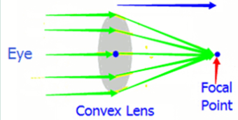
Convex lenses are used in things like telescopes and binoculars to bring distant light rays to a focus point where they can be transposed to paper or the computer or viewed directly.
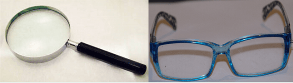
Two common lenses are the hand lens and eye glasses. A simple desk mounted hand lens can be used to magnify objects up to about 30 times their original size.
Make a simple water lens.
1. Take an old newspaper or magazine and remove a page.
2. Lay a small piece of cellophane, cling film, or clear plastic on top of the sheet. Place it over the paper and level it out so there are no creases in it.
3. Use an eye dropper, pipette, syringe place a single, small drop of water on top of the cling plastic film.
4. Look at the newsprint and you should be able to see that the water drop (which has a curved upper edge and a flat lower edge) magnifies the words.
5. Repeat the above with a small amount of sugar or salt in the water. Now look at what happens?
6. What happens if you make the water drop bigger or smaller?
7. What happens if you move the plastic away from the paper or the plastic has a slight convex sag in it?
This is how all great scientists began using a little initiative and what equipment is available and played around or experimented.

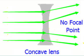
A concave lens is the opposite of a convex lens. The surfaces curve inwards and is also called a diverging lens because it makes parallel light rays passing through it bend outward. There is no focal point as such.
The focal point may be on the receiving side of the concave lens.
A concave lens makes light rays diverge (spread out).
Concave lenses are used in things like TV projectors to make light rays spread out into the distance. In a flashlight, it’s easier to do this job with a mirror, which usually weighs much less than a lens and is cheaper to manufacture as well.
Compound Lens – The optical or Light Microscope:
A lens that uses two or more simple lenses; often by combining convex and concave lenses, is called a compound lens. Take for example the camera which combines the different lens to give different results. Modern cameras also change the focal length. There’s a simple measurement that tells you how powerful a lens is and it’s known as the focal length. The focal length of a lens is the distance from the center of the lens to the point at which it focuses light rays. The shorter the focal length is the more powerful the lens becomes. The microscope is another form of using compound lenses.
Standard optical microscopes are limited in the following ways:
* Diffraction limits resolution to about 0.2 millimeters,
* Only dark or strongly refracting objects can be imaged,
* Light from outside sources reduces the clarity of the image.
To overcome these limitations, several different techniques of microscopy; the science of the use of microscopes, have been invented which include the following specific Apparatus and their Functions
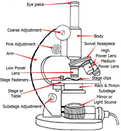
* The eyepiece contains a lens which magnifies the image while the body tube separates the two lenses.
* The revolving nosepiece allows the user to rotate between the three different lenses.
* The coarse adjustment knob moves the tube for focusing the weak magnification lens; the fine adjustment knob moves the tube for focusing the strong magnification lens.
* The stage supports the slide being viewed under the lens.
* The aperture is a hole in the stage for light to pass through.
* The diaphragm controls the amount of light entering the stage through the aperture which is located in the rack and pinion substage.
* Stage clips are for holing the specimen slides.
The microscopes allowed serious advances in science especially in the field of medicine. This only became possible with the increased knowledge of microscopic life forms which made bacterial microbes both visible and real. Prior to microscopes, people attributed diseases to harmful spiritual means. With the invention of the optical microscope having a high numerical aperture and using oil immersion, the best possible resolution is 200nm which corresponds to a magnification of around 800 times (800x). With the advent of high quality immersion oils, better lens manufacture the maximum usable magnification increased to 2000x.
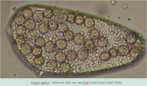
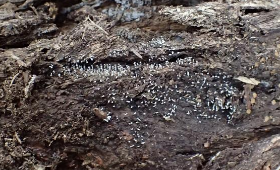
Fuligo septica x1 – andi Mellis

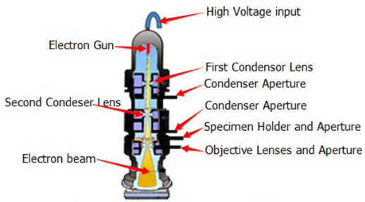
Electron microscope:
An electron microscope is a microscope that uses accelerated electrons as a source of illumination. Because the wavelength of an electron can be up to 100,000 times shorter than that of visible light photons, the electron microscope has a higher resolving power than a light microscope and can reveal the structure of much smaller organisms. A transmission electron microscope can achieve better than 50pm resolution and magnifications of up to about 10,000,000x.
The transmission electron microscope (TEM) uses electrostatic and electromagnetic lenses to control the electron beam and focus it to form an image. These electron optical lenses are similar to the glass lenses of an optical light microscope.
Electron microscopes are used to investigate the ultrastructure of a wide range of biological and inorganic specimens including microorganisms, cells, large molecules, biopsy samples, metals, and crystals. Industrially, the electron microscope is often used for quality control and failure analysis. Modern electron microscopes produce electron micrographs, using specialized digital cameras or frame grabbers to capture the image.
Resolution of the TEM is limited primarily by spherical aberration, however the new generation of aberration correctors has been able to partially overcome spherical aberration to increase resolution. Hardware correction of spherical aberration for the high-resolution transmission electron microscopy (HRTEM) has allowed the production of images with resolution below 0.5 angstrom (50 picometres)[1] and magnifications above 50 million times. The ability to determine the positions of atoms within materials has made the HRTEM an important tool for nano-technologies research and development.
Unlike the TEM, where electrons of the high voltage beam carry the image of the specimen, the electron beam of the scanning electron microscope (SEM) does not at any particular time carry a complete image of the specimen. The SEM produces images by probing the specimen with a focused electron beam that is scanned across a rectangular area of the specimen. When the electron beam interacts with the specimen, it loses energy by a variety of mechanisms. The lost energy is converted into alternative forms such as heat, emission of low-energy secondary electrons and high-energy backscattered electrons, light emission (cathodoluminescence) or X-ray emission, all of which provide signals carrying information about the properties of the specimen surface, such as its topography and composition. The image displayed by an SEM maps the varying intensity of any of these signals into the image in a position corresponding to the position of the beam on the specimen when the signal was generated.
The image resolution of an SEM is at least an order of magnitude poorer than that of a TEM. However, because the SEM image relies on surface processes rather than transmission, it is able to image bulk samples up to many centimeters in size and has a great depth of field, and so can produce images that are good representations of the three-dimensional shape of the sample. Another advantage of SEM is its variety called environmental scanning electron microscope (ESEM) can produce images of sufficient quality and resolution with the samples being wet or contained in low vacuum or gas. This greatly facilitates imaging biological samples that are unstable in the high vacuum of conventional electron microscopes.
In their most common configurations, electron microscopes produce images with a single brightness value per pixel, with the results usually rendered in grey. However, often these images are then colorized through the use of feature-detection software, or simply by hand-editing using a graphics editor. This is usually for aesthetic effect or for clarifying structure, and generally does not add information about the specimen.
The Reflection Electron Microscope (REM) like the TEM uses an electron beam is incident, on a surface but instead of using the transmission (TEM) or secondary electrons (SEM), the reflected beam of elastically scattered electrons are detected. Materials to be viewed under an electron microscope may require
Tem, SEM and REM are used where higher magnification is sought particularly important in the nano sciences.
Electrical:
Semiconductor and data storage
Defect analysis
Failure analysis
Biology and life sciences:
Diagnostic electron microscopy
Cryobiology
Protein localization
Electron tomography
Cryo-electron microscopy
Toxicology
Biological production and viral load monitoring
Particle analysis
Pharmaceutical QC
Structural biology
3D tissue imaging
Virology
Vitrification
Materials research
Electron beam-induced deposition
Materials qualification
Medical research
Nano:
Nanoprototyping
Nanometrology
Device testing and characterization
Industry & Fornsics:
High-resolution imaging
2D & 3D micro-characterization
Macro sample to nanometer metrology
Particle detection and characterization
Direct beam-writing fabrication
Dynamic materials experiments
Sample preparation
Mining:
Mineral liberation analysis
Chemical / Petrochemical
Fractography and failure analysis
Back to Microbiology and evolution it was the sheer dominance of microorganisms in both biomass and biodiversity that led to specie variation with the dominance being played out within the prokaryotes. Even today micro organisms are the dominant species on Earth and far out number the macro organisms in both specie diversity and biomass.
These organisms can only be seen under the microscope but despite their size these micro organisms, or microbes for short, have a massive impact on our lives. It has been estimated that there are 5 million trillion, trillion, microbial cells on Earth. The total amount of carbon in these cells is equivalent to that of all of the plants and animals on the planet. They collectively constitute the largest mass of living material on earth and play a critical role in shaping the environment that we live in. Humans, plants and animals are intimately tied to the activities of microbes which recycle key nutrients and degrade organic matter.
Microbes have existed on Earth for billions of years and were here long before plant and animal life began. For the majority of the Earth’s 4.5 billion year history, life on Earth was exclusively microbial. Microbial cells first appeared between 3.8 and 3.9 billion years ago. The fossilised remains of these early bacteria can be detected in stromatolites or rock like accumulated conglomerates of microbial mats and trapped sediment. When the Earth first formed there was no free oxygen present and only bacteria which could grow without oxygen could thrive. Eventually a group of bacteria know as cyanobacteria evolved which were able to photosynthesise and began producing oxygen as a waste by product. At this point the long process of oxygenating the world began, starting the slow, gradual process of the evolution from anaerobic organisms to aerobic organisms including animals and plants.
Now that we are able to see much smaller organisms the science of microbiology began to flourish. Single celled plants and animals, individual cells along with viruses are now able to be studied in far greater detail.
It wasn’t until the invention of the microscope that the study of microbes became its own science. The microscope was one of key discoveries along with improved laboratory techniques and an understanding of how microbes function and interact with their environment that advanced the science of microbiology.
Microbiologists focus on how microbes function, reproduce and interact with their environment. They also investigate the practical applications of microbes in medicine, agriculture, industrial processes and other areas.
Many of the most important discoveries by microbiologists have been in the field of techniques for making indirect observations of microbes and their activities. Since the first observation of microbes in a single droplet of lake water using of the microscope; in the late seventeenth century, microbiologists have been fascinated with the unseen world around us. Other techniques microbiologists studied included the isolation and growing microbes in the laboratory leading to sterilization procedures.
The early history of microbiology focused on two important areas those being fermentation and medicine. The medical discoveries, in particular, have received more recognition. Important medical discoveries include the development of vaccines and the inoculation of societies against harmful microbes. More recently, scientists learned how to study and manipulate the genetic information of microbes. This field of microbial genetics continues to have important potential for medicine, agriculture and industrial applications. This has been most controversial in genetically modified foods where human health is directly affected.
Microbe Theories
In the fourth century BC, Aristotle proposed that in addition to sexual and asexual reproduction, organisms could appear suddenly from inanimate matter. He called this theory, “spontaneous generation”. This idea persisted for quite some time, especially in connection to “microbes.” Louis Pasteur proved through a series of carefully designed experiments that bacteria did not grow from nothing, but came from other bacteria. At about the same time, scientists were exploring the specifics of how microbes cause disease, known as the germ theory of disease.
Key Personnel and Institutions of Microbiology
One of the first was microbiologists was a Dutch draperyist; Antoni van Leeuwenhoek, who used an early microscope to view pond water, thus initiating the study of microbes. Louis Pasteur advanced microbiology by disproving Aristotle’s theory of spontaneous generation and developing methods of vaccinating against viruses. Robert Koch, who promoted growing single types of microbes on agar material, provided the final evidence showing that microbes caused disease. The work of Alexander Fleming in the early twentieth century with the antibiotic penicillin signaled the start of a new era in medical microbiology.
Much of medical microbiology involves treating sick people, as well as preventing diseases from spreading throughout a population. Government agencies, such as the World Health Agency and the Centers for Disease Control, are heavily involved in helping people stay healthy and dealing with outbreaks of disease. This includes monitoring the spread of infections like influenza and AIDS, as well as identifying dangerous new microbes, such as ebola and bacteria resistant to antibiotics.
Food for Thought – The good guys:
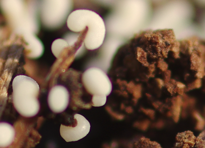
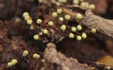
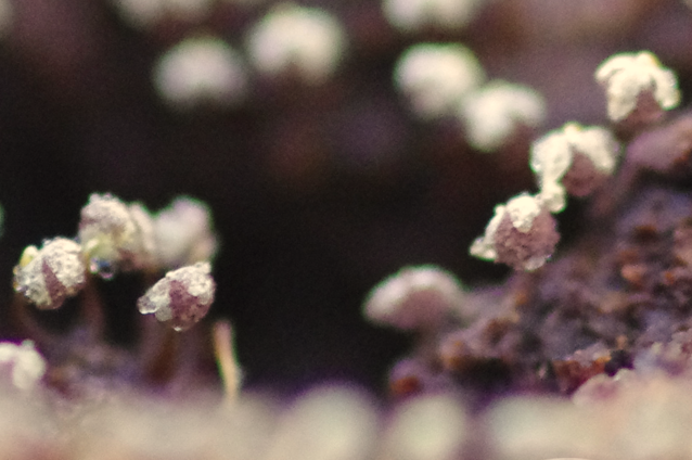
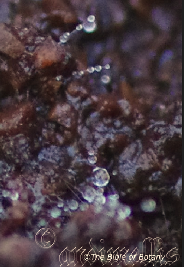
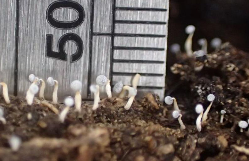
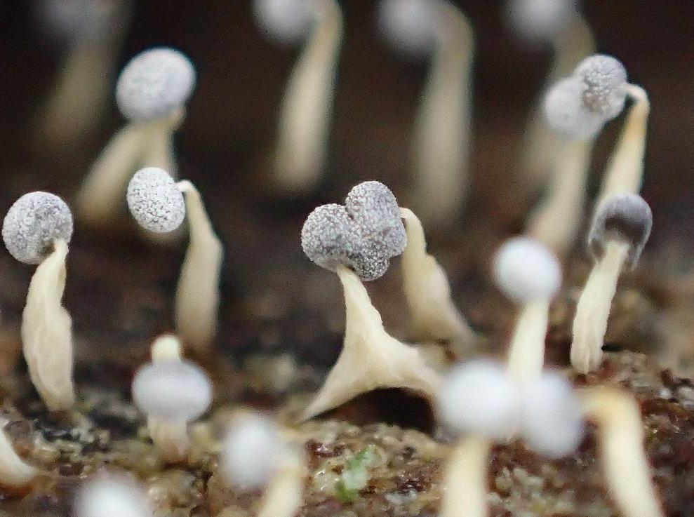
Because of great specie diversity in microbes living on the planet which includes bacteria, fungi, protozoa, algae, parasites and viruses scientists can have a great diversity bin careers. Rather than focusing on one type of microbe, microbiologists can also study broad topics within microbiology including the structure, genetics or physiology of microbes, how microbes interact with their ecosystems, such as a pond or the human body. Scientists may also study how microbes have changed over time, in what is known as evolutionary microbiology. Below are three Physarum species at different stages of fruiting development and magnification.
Physarum specie are wood decomposing fungi. Physarum paniceum x5 on Brachychiton aceriifolius – andi Mellis
Physarum paniceum x15 – andi Mellis
Physarum sp. x7.5 prior to spore maturing – andi Mellis
Physarum sp. x15 at point of spore release – andi Mellis
Physarum sp. x30 showing gleba and the remains following spore release – andi Mellis
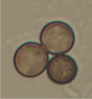
Physarum nucleatum x1 as seen by the human eye. – https://fungimap.biodiv.tw/pages/952
Physarum nucleatum x10 – https://fungimap.biodiv.tw/pages/952
Physarum nucleatum x40 – https://fungimap.biodiv.tw/pages/952
Physarum nucleatum x900 – Teresa & John van der Huel South Coast NSW.
Industrial microbiology involves the use of microbes for food processes such as fermentation. One area of fermentation that people do not think of is wastewater treatment. Yes the primary, secondary and tertiary treatment of our waste water all comes down to the good old microbe. Take a close look inside your septic tank not too close though and you will see with the use of a microscope millions of the little critters chewing away, lapping up another piece of human waste. Ah yes microbes.
Industrial microbiology involves the use of microbes for food processes such as fermentation. One area of fermentation that people do not think of is wastewater treatment. Yes the primary, secondary and tertiary treatment of our waste water all comes down to the good old microbe. Take a close look inside your septic tank not too close though and you will see with the use of a microscope millions of the little critters chewing away, lapping up another piece of human waste. Ah yes microbes.
Babies are colonised by bacteria immediately after birth. It has been estimated that the average person is colonised by 200 trillion bacteria, comprising at least 1,000 different specie. The bacteria that call the human body home are often essential for our health and well being. Our intestines contain about 100 trillion bacteria and collectively they make up 60mm of the dry weight of faeces. Your intestinal bacteria are essential in helping you to digest your food, they provide you with essential vitamins such as vitamin K and biotin and they help to prevent the growth of harmful pathogenic bacteria. The surface of our skin is also home to millions of friendly bacteria which crowd out potential pathogens and prevent them from growing. One bacterium which is abundant on the skin is Staphylococcus epidermidis which produces chemicals called bacteriocins that kill pathogenic bacteria.
Well maybe I will have to give up my daily shower for the sake of Staphylococcus epidermidis.
Friendly bacteria are teaming up your nose but many of these bacteria also carry health warnings. Neisseria meningnitidis which causes meningitis, lives in the noses of millions of people without causing disease, but if the immune system becomes weakened through ill health then this bacteria can, almost by accident, cause disease which may result in the death of the human that has become its home. Well they better not get up my nose!
Something probably a little more interesting and a little less disgusting are all those microbes we eat on a daily basis. Yes some of these specie are responsible in the processes other kinds of fermentation like the production of alcohol, vinegar, dairy products like yoghurt or different yeast in the production of our daily bread and of course the manufacturing of that famous Australian spread vegemite.
Microorganisms don’t stop there they also provide us with pleasure! Microbes are involved in making chocolate? The cocoa pods are split open and their contents of 20 to 30 bitter seeds in a sweet sticky pulp are heaped together and covered with banana skins and naturally fermented for 7 days. Over 30 different types of bacteria are involved in this process, along with yeasts and moulds. While in this heap, the sticky pulp becomes a turbid chocolate coloured broth which gives the cacao seeds both their characteristic chocolate flavour and colour. Good old microbes.
Salt loving bacteria, like those found in salt flats, play a key role in the production of Thai fish sauce and Japanese soy sauce. Salt loving bacteria are also important in the production of cured meats and sausages such as salami.
And for those who love Chinese cooking well it also depends heavily on microbes which are essential in the production of black bean and yellow bean sauces. Then let’s not forget smelly tofu or any tofu for that fact are the conversion of soy milk into tofu using you guessed it microbes.
I cannot leave without mentioning one significant little bacteria. It is used in the industrial production of amino acids. Corynebacterium glutamicum is one of the most important bacterial species to humans with an annual production of more than two million tons of amino acids, mainly in the forms of L-glutamate and L-lysine.
Without microbes our culinary repertoire would be smaller and our diets extremely bland.
So apart from your usual intake of fruit and vegetables how many microorganisms did you consume today?
Food for Thought – The bad guys:
The interaction between microbes and food is not always the best as it can foul the food causing foul odours, bitterness in taste and worse still diarrhoea. Foods which have passed their sell by date may not be obviously contaminated with bacteria. The sell by dates are based on the amount of time it takes for the numbers of bacteria to reach a level of micro bacterial contamination where the chances of food poisoning begin to increase. The bacterium Campylobacter jejunii is the most common cause of food poisoning in the UK.
In 2010 the Food Standards Agency estimated that 65mm of all fresh chickens sold in the UK were contaminated with Campylobacter jejunii. Cooking kills this bacterium but it is still responsible for approximately 300,000 cases of food poisoning in England and Wales each year. For most healthy people, food poisoning, although very unpleasant, is not life threatening. However for both babies and elderly people food poisoning can be extremely serious as it can cause severe dehydration and kidney failure. These bacteria also affect the good bacteria in the gut which can further exasperate any problems.
Escherichia coli and Staphylococcus aureus are 2 potentially harmful bacteria that are commonly found in water, food and on clean animal skins. In mild doses the body expels them daily but in large numbers can be the cause of different food poisonings with wild to severe vomiting and Diarrhoea. Human perspiration harbours and supports Staphylococcus epidermidis in which their excrements contain natural antimicrobial proteins.
Protectors of the Environment:
Many microbes like plants and scavengers act as guardians of our planet ensuring that key minerals, such as carbon and nitrogen, are constantly recycled back into the earth. Even though the Earth is now populated with green plants, microbes still play a crucial role in oxygenating the atmosphere and collectively they carry out more photosynthesis than plants. Can you believe that? Microbes degrade dead organic matter, converting the organic carbon in their bodies back into carbon dioxide and soluble carbonic acid for use by other organisms.
Microbes also play a key role in the nitrogen cycle. Bacteria in the soil attach themselves to the roots of plants like the Fabaceae and Casuarinaceae and convert atmospheric nitrogen into nitrates for the use by the plants or are free in the soil where they convert soil nitrogen in the soil to nitrates. Nitrates are an essential plant nutrient as they are essential in the production of proteins. Plants are the producers of food for live other organisms like animals. The nitrogen locked in plant and animal proteins is then degraded into nitrates by microbes and eventually converted back into nitrogen by denitrifying bacteria.
Compost heaps are phenomenal exhibitions of microbes effectively transforming insoluble organic matter into soluble organic matter. A mixture of garden waste, household waste and animal waste is rapidly decomposed by fungi and bacteria. Without the recycling power of microbes dead vegetation, carcasses and food waste would start piling up around us! In the United Kingdom 6.7 million tonnes of food waste is discarded every year. Imagine the implications if this waste just sat around to accumulate?
Microbes are Everywhere:
Microbiologists traditionally relied on culture, staining, and microscopy. Less than 1mm of the microorganisms present in our environments can be cultured in isolation using current means.Microbiologists often rely on extraction or detection of nucleic acid, either DNA or RNA sequences.
Microbiologists have discovered that microbes can be found in every niche on Earth. Microbes are an incredibly diverse group of organisms and can grow in extreme environments that no living macro organisms can tolerate. Bacteria have been found to thrive in volcanic hot springs, where temperatures typically reach near boiling point even those several kilometres beneath the ocean along hot fissures. At the other extreme, living bacteria have also been discovered in Antarctic deserts, where temperatures range from -15 to -30°C. Bacteria can also thrive in salt flats, pools of saturated brine, where salt concentrations range from 120 to 230 grams per litre. Bacteria which live happily in these inhospitable environments have been termed ‘extremophiles,’ from Latin extremus meaning “extreme” and Greek philiā meaning “love or lover”.
In addition to being a biological curiosity bacteria which grow in these extremely hostile conditions have proven to be a rich source of enzymes for the biotechnology industry. Fat decomposing and protein decomposing enzymes from bacteria isolated from hot springs have been used to make ‘biological washing powders’. Unlike equivalent enzymes from ‘ordinary’ bacteria these function efficiently at the high temperatures typically used for doing the laundry. Clearing up oil spills that have occurred in cold oceanic environments, the production of ice cream and artificial snow have also benefited from enzymes, produced by bacteria that thrive in near zero temperatures.
The study of infectious disease is another important branch of microbiology. At the beginning of the 20th century infectious diseases, caused by microbial pathogens, were the major cause of death. Large numbers of children and the elderly succumbed to diseases such as tuberculosis, diphtheria and pneumonia. At this time microbiologists had little idea about how diseases were spread, or how they could be controlled, so epidemics flourished. The “Spanish” influenza pandemic of 1918–1919, caused around 50 million deaths worldwide, more than the total number of deaths recorded in World War One. Diarrhoeal disease was also common since people regularly ate contaminated food and drank contaminated water.
Microbiologists are therefore concerned with developing antibiotics and vaccines to protect the population from infectious disease. The discovery of penicillin by Alexander Flemming has saved the lives of many millions of people. The development of vaccines which protect against, diphtheria and pneumonia dramatically reduced the number of childhood deaths caused by these diseases. Children in developed countries are also routinely vaccinated against common viral infections such as measles, mumps, rubella and polio. As a direct result of the efforts of microbiologists, smallpox, once an insidious disease, is now officially extinct on the planet. However a vaccine against HIV, which in 2009 was reported to infect approximately 33.3 million people around the world still eludes us.
Viruses have been invariably classified as organisms at times, as they have been considered either as very simple micro organisms or very complex protein molecules.
As an application of microbiology, medical microbiology is often introduced with medical principles of immunology as microbiology and immunology. Otherwise, microbiology, virology, and immunology as basic sciences have greatly exceeded the medical variants, applied sciences.
The branches of microbiology can be classified into pure and applied sciences. Microbiology can be also classified based on taxonomy, in the cases of bacteriology, mycology, protozoology, and phycology. There is considerable overlap between the specific branches of microbiology with each other and with other disciplines, and certain aspects of these branches can extend beyond the traditional scope of microbiology.
For more on microbiology see water and Land Resource Management.
Further Comments from Members:
All information is included in good faith and has been thoroughly researched prior to printing. The website or the author does not warrant or guarantee the accuracy of any information on these pages, nor does the website or the author accept any responsibility for any loss arising from the use of the information found within. The views and opinions are strictly those of the author or those members who chose to actively participate in the contents herein.
“Hi reader, it seems you use The Bible of Botany a lot. That’s great as we have great pleasure in bringing it to you! It’s a little awkward for us to ask, but our first aim is to purchase land approximately 1,600 hectares to link several parcels of N.P. into one at The Pinnacles NSW Australia, but we need your help. We’re not salespeople. We’re amateur botanists who have dedicated over 30 years to saving the environment in a practical way. We depend on donations to reach our goal. If you donate just $5, the price of your coffee this Sunday, We can help to keep the planet alive in a real way and continue to bring you regular updates and features on Australian plants all in one Botanical Bible. Any support is greatly appreciated. Thank you.”
In the spirit of reconciliation we acknowledge the Bundjalung, Gumbaynggirr and Yaegl and all aboriginal nations throughout Australia and their connections to land, sea and community. We pay our respect to their Elders past, present and future for the pleasures we have gained.